Early Development of the Brain
Introduction
The central nervous system, and the brain, in particular, are incredibly complex, with a variety of systems, brain regions, and subregions, which can be hard to distinguish.
Starting from an embryologic perspective allows you to understand more easily how the parts relate to each other. The embryonic nervous system begins as a very simple structure—essentially just a straight line, which then gets increasingly complex. Looking at the development of the nervous system with a couple of early snapshots makes it easier to understand the whole complex system.
During prenatal development, there are three stages of physical growth: germinal period, embryonic period, and the fetal period.
First, during the germinal period, the developing organism is called a zygote. This period occurs from fertilization of the egg by sperm until two weeks after fertilization.
Second, during the embryonic period, the zygote is now called an embryo. This period occurs from two weeks after fertilization until eight weeks after fertilization. During this time, major organs and bodily structures begin to form.
Third, and finally, during the fetal period, the embryo is now called a fetus. This period occurs from eight weeks after fertilization until birth. During this time, bodily structures are refined, the fetus grows in length and width and accumulates fat, and the brain continues to develop.
In this activity, you will learn about the stages of brain development. These stages include the following steps:
- Formation of the neural plate
- Proliferation of brain cells
- Migration of brain cells
- Differentiation of progenitor cells
- Development of appendages
- Pruning and refinement of cells
“Anatomy and Physiology: Chapter 13, Section 1: The Embryologic Perspective” by OpenStax. Retrieved from: https://opentextbc.ca/anatomyandphysiology/chapter/13-1-the-embryologic-perspective/. Licensed under CC BY.
Stages of Brain Development
Formation of the Neural Plate
To begin, a sperm cell and an egg cell fuse to become a fertilized egg. The fertilized egg cell, or zygote, starts dividing to generate the cells that make up an entire organism.
During approximately the second week after fertilization, a neural plate forms on the dorsal portion of the embryo.
As the zygote develops, a layer of tissue is induced into forming a neural plate. The cells then begin to change shape, causing the tissue to buckle and fold inward. A neural groove forms, which becomes visible as a line along the dorsal surface of the embryo. The ridge-like edge on either side of the neural groove is referred to as the neural fold. As the neural folds come together and converge, the underlying structure forms into a tube called the neural tube.
To summarize, during approximately the third week after fertilization, this neural plate pinches to form the neural tube, which eventually develops into the central nervous system (the brain and the spinal cord). The most interior portion of the neural tube becomes the ventricles, which are filled with cerebrospinal fluid to provide cushioned protection.
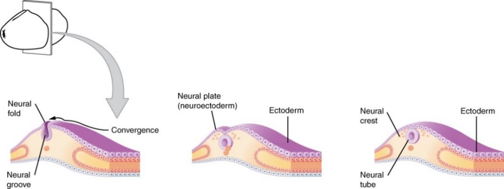
The image shows the early embryonic development of the nervous system in three panels. On the left, a cross-section of tissue is shown, with the neural fold, neural groove, and convergence shown. In the middle, the neural plate has formed and is now called the neuroectoderm and is separate from the ectoderm. On the right, the neural crest, neural tube, and ectoderm are labelled.
“Anatomy and Physiology: Chapter 13, Section 1: The Embryologic Perspective.” by OpenStax. Retrieved from: https://opentextbc.ca/anatomyandphysiology/chapter/13-1-the-embryologic-perspective/. Licensed under CC BY.
Proliferation of Brain Cells
Once the neural tube has developed, progenitor cells in the ventricular zone continue to divide. These progenitor cells have the potential to become one of many different types of cells – they are currently undifferentiated. Indeed, they are similar to stem cells, but have more constrained options. The ventricular zone is the layer of cells that lines the ventricles, the most interior portion of the neural tube.
Migration of Brain Cells
Although progenitor cells continue to divide, they cannot all stay in the same location – the most interior part of the neural tube. Instead, many of these cells are destined to be on the exterior of the brain.
Critically, the cortex (the wrinkly portion of the brain) develops in an “inside out” matter. Indeed, although cells are formed in the interior, many must migrate from the inner layer to an outer one.
To accomplish this movement, these cells migrate on radial glia, supporting cells that essentially provide “railroad tracks” for these migrating progenitor cells.
Differentiation of Progenitor Cells
As mentioned, progenitor cells are undifferentiated, with the potential to become many different types of cells. Once these progenitor cells reach their destination via migration on the radial glia, they can differentiate.
In particular, these progenitor cells undergo either neurogenesis or gliogenesis. In neurogenesis, new neurons are generated from progenitor cells. In gliogenesis, new glia are generated from progenitor cells.
How do progenitor cells know what to become? These cells differentiate in response to inducing factors, which are chemical signals that determine the fate of a cell. A chemical signal, or inducer, will activate certain genes. Due to this gene expression, specific proteins are synthesized. Do you remember your biology background? The central dogma of biology is the process of DNA RNA proteins. These proteins are the building blocks of our cells and our bodies.
Incredibly, during embryonic development, approximately 250,000 neural cells (neurons and glia) are generated per minute! As you will soon discover, not all of these cells will survive. But, for now, a massive number of cells are generated to provide the foundation for the central nervous system.
Development of Appendages
Now that the cells have migrated and differentiated – that is, they have specialized into either a neuron or glia (e.g. oligodendrocyte, astrocyte) – they must develop the necessary tools for cellular communication.
During this stage of development, neural cells develop axons and dendrites, in order to send and receive information, respectively. These appendages develop with the help of growth cones, swellings at the tips of axons and dendrites that guide the appendage to the target location. To be even more precise, these growth cones are guided by genetic codes and chemical signals.
Once an axon nears another cell, a synapse is formed. Recall that a synapse is the gap between one cell and another, in which communication occurs. The most common synapse is that between an axon and a dendritic site, which requires synaptic vesicles to form in the axon terminal as well as receptors to form at the point of contact on the dendrite.
Pruning and Refinement of Cells
As you will recall from the earlier foreshadowing, not all cells will survive. Indeed, the brain should be efficient, meaning that it does not have cells that do not serve a purpose.
How does the brain accomplish its pruning and refinement in order to achieve efficiency? Cells produce neurotrophins, which are chemicals that promote cell survival. For example, a common neurotrophin is brain-derived neurotrophic factor (BDNF).
Cells and synapses that receive sufficient neurotrophins will survive. All others will undergo apoptosis, or programmed / planned cell death. During this time, approximately 100,000 cells die per minute!
Donald Hebb coined the phrase, “Cells that fire together, wire together.” Indeed, if a cell causes a target cell to fire, then that targeted cell will release neurotrophins, resulting in its “tag” for survival. However, cells that are not caused to fire will not survive. This same concept is true not only for the cells themselves, but also for the specific synapses. If some synapses are not being used, they will degrade.
Consider this metaphor: as an urban developer, you have built several neighborhoods and streets to connect them. However, not all neighborhoods and streets will be used. You assess their use and “tag” the necessary areas with signs, saying “in use.” All non-tagged areas will be demolished, so that they do not use up important resources and tax dollars – street cleaning, waste pick-up, plumbing, electricity, etc. Indeed, in early development, the brain is similar, operating under a “use it or lose it” axiom.
So how does this manifest in the behavioural world, one that we can see? To answer this question, you must understand the concept of a critical period, which is the period of time during development in which a particular experience is influential. After the critical period expires, experience has little to no effect on structure.
Consider two examples in kittens that are still developing their visual system.
Example #1: Scientists can cover one eye. In doing this manipulation, one eye will be receiving input and sending signals to the brain, whereas the other eye will not be used. After the critical period has transpired, the unused eye will be “blind.” Although there is no damage to the eye, its cells and connections were not used, and so they were not kept.
Example #2: Scientists can place the kitten in a circular arena, in which there are no vertical lines. In doing this manipulation, the kitten will not receive any input requiring the processing of vertical lines. After the critical period has transpired, the kitten is unable to perceive vertical lines, and would be at risk for running into the legs of tables and chairs. Again, although there is no damage to the eye, the cells and connections necessary for the processing of vertical lines were not used, and so they were not kept.
Tanaka, S., Tani, T., Ribot, J., O’Hashi, K., & Imamura, K. (2009). A Postnatal Critical Period for Orientation Plasticity in the Cat Visual Cortex. PLoS ONE, 4(4), e5380. Retrieved from http://doi.org/10.1371/journal.pone.0005380.
Voss, P. (2013). Sensitive and critical periods in visual sensory deprivation. Frontiers in Psychology, 4, 664. Retrieved from http://doi.org/10.3389/fpsyg.2013.00664.
Stages of Brain Development – Video
Neuroscientifically Challenged. (2014, November 7). 2-minute neuroscience: Early Neurodevelopment. [Video File]. Retrieved from: https://www.youtube.com/watch?v=Tp25wrm-AoA. Standard YouTube License.
Think Psych. (2016, September 12). Prenatal brain development. [Video File]. Retrieved from https://www.youtube.com/watch?v=XdN9i_ZWGho. Standard YouTube license.
BMEN 5411 Neural Engineering. (2015, June 15). Cell migration: Refinement. [Video]. Retrieved from https://www.youtube.com/watch?v=JkPyShERBjE. Standard YouTube license.
Summary
The central nervous system (brain and spinal cord) begins developing approximately two weeks after fertilization, during the germinal period of prenatal development.
Critical stages of brain development include the following:
- Formation of the neural plate = neural plate becomes neural tube with ventricular zone
- Proliferation of brain cells = progenitor cells divide in the ventricular zone
- Migration of brain cells = progenitor cells move, via radial glia, in an “inside out” manner
- Differentiation of progenitor cells = undifferentiated cells undergo neurogenesis or gliogenesis
- Development of appendages = cells develop axons and dendrites via growth cones and inducing factors, resulting in synapses
- Pruning and refinement of cells = only used cells and synapses survive due to neurotrophic factors
These processes occur during prenatal development, which consists of three stages of physical growth: germinal period of the zygote, embryonic period of the embryo, and fetal period of the fetus.