Neural Communication
Introduction
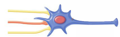
The nervous system is exactly that – a system of nerves. This network of special cells sends and receives messages. These cells, or neurons, are special because they send and receive messages in a different way than the cells of the body.
The nervous system has three basic functions:
1. To receive input through senses (sight, sound, taste, smell, touch)
2. To process information through cognition (attention, perception, learning)
3. To respond to information through action (movement)
It might be intuitive, then, to think that a system with such requirements would be built out of cells that have such abilities.
Indeed, neurons have three basic functions:
1. To receive signals from other neurons
2. To process signals via summation
3. To send signals to other neurons
Consider your own experience participating in a group discussion. As you sit amidst your peers, you are listening to information. If you are stimulated by enough information, you will make a decision and speak up.
Neurons operate in a comparable way: they receive information in the form of chemical messengers (neurotransmitters) from other neurons – they may be receiving many different types of chemicals from many different neurons. They summate (or add together) these messages and, if they are stimulated enough, they will send their own electrical signal, resulting in the release of neurotransmitters.
Neural Communication
Electrochemical Signalling
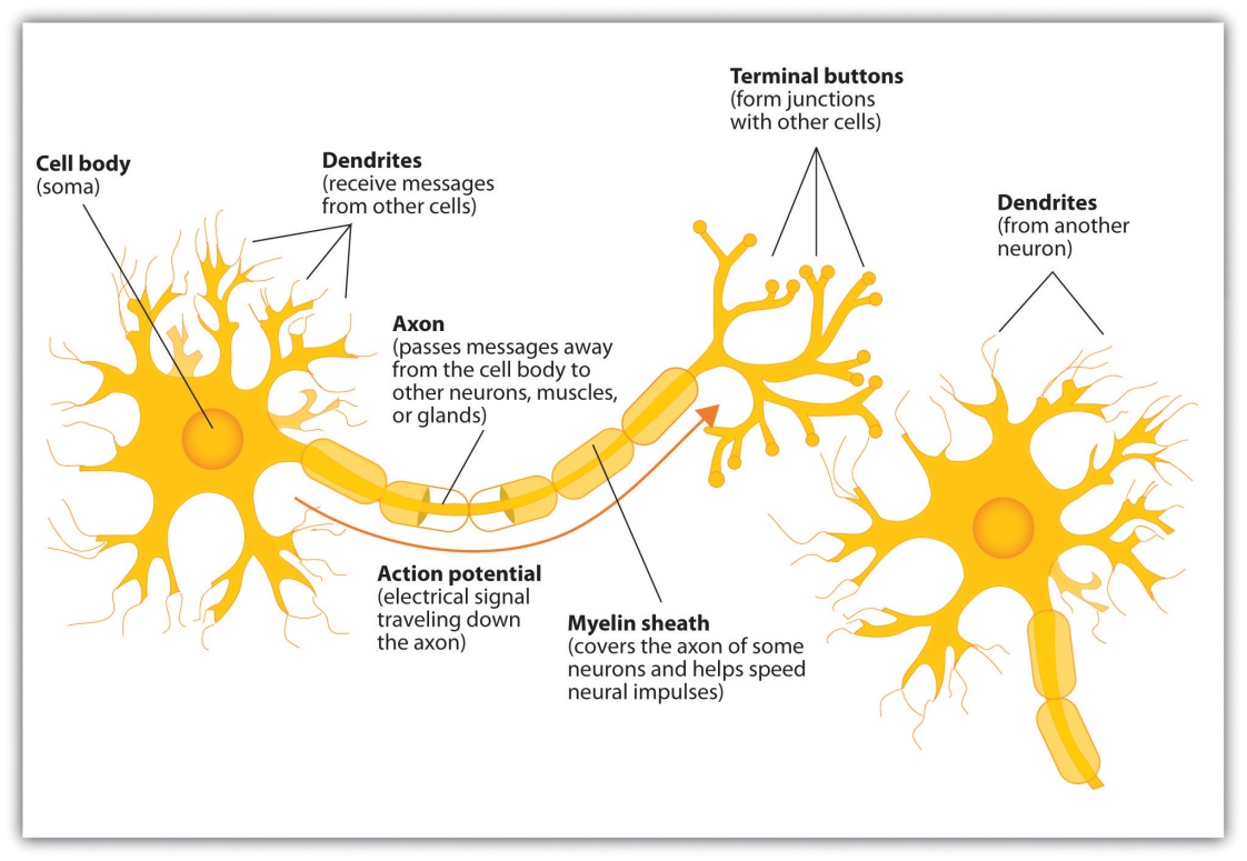
The nervous system operates using an electrochemical process. An electrical charge moves through the neuron itself and chemicals are used to transmit information between neurons. Within the neuron, when a signal is received by the dendrites, it is transmitted to the soma in the form of an electrical signal, and, if the signal is strong enough, it may then be passed on to the axon and then to the terminal buttons. If the signal reaches the terminal buttons, they are signalled to emit chemicals known as neurotransmitters, which communicate with other neurons across the spaces between the cells, known as synapses.
“Introduction to Psychology: Chapter 3, Section 1: The Neuron Is the Building Block of the Nervous System” by the University of Minnesota Retrieved from http://open.lib.umn.edu/intropsyc/chapter/3-1-the-neuron-is-the-building-block-of-the-nervous-system/#stangor-ch03_s01_s01_n01Licensed under CC BY-NC-SA.
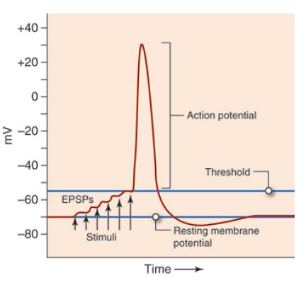
The electrical signal moves through the neuron as a result of changes in the electrical charge of the axon. Normally, the axon remains in the resting potential, a state in which the interior of the neuron contains a greater number of negatively charged ions than does the area outside the cell. When the segment of the axon that is closest to the cell body is stimulated by an electrical signal from the dendrites, and if this electrical signal is strong enough that it passes a certain level or threshold, the cell membrane in this first segment opens its gates, allowing positively charged sodium ions that were previously kept out to enter. This change in electrical charge that occurs in a neuron when a nerve impulse is transmitted is known as the action potential. Once the action potential occurs, the number of positive ions exceeds the number of negative ions in this segment, and the segment temporarily becomes positively charged.
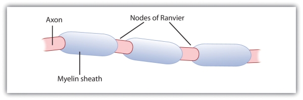
The axon is segmented by a series of breaks between the sausage-like segments of the myelin sheath. Each of these gaps is a node of Ranvier. The electrical charge moves down the axon from segment to segment, in a set of small jumps, moving from node to node. Indeed, this process is called saltatory conduction (Latin, saltare – to jump). When the action potential occurs in the first segment of the axon, it quickly creates a similar change in the next segment, which then stimulates the next segment, and so forth, as the positive electrical impulse continues all the way down to the end of the axon. As each new segment becomes positive, the membrane in the prior segment closes up again, and the segment returns to its negative resting potential. In this way, the action potential is transmitted along the axon toward the terminal buttons. The entire response along the length of the axon is very fast—it can happen up to 1,000 times each second!
“Introduction to Psychology: Unit 4, Module 5: Neurons Communicate Using Electrochemical Processes” by Open Learning Initiative. Retrieved from https://canvas.instructure.com/courses/773049/pages/stangor-3-dot-1-the-neuron-is-the-building-block-of-the-nervous-system. Licensed under CC BY-NC-SA.
The myelin sheath wraps around the axon, but also leaves small gaps called the nodes of Ranvier. The action potential jumps from node to node as it travels down the axon.
An important aspect of the action potential is that it follows the all-or-none principle, meaning that it operates in an all-or-nothing manner. What this means is that the neuron either fires completely, such that the action potential moves all the way down the axon, or it does not fire at all. Thus, neurons can provide more energy to the neurons down the line by firing faster but not by firing more strongly. Furthermore, the neuron is prevented from repeated firing by the presence of a refractory period—a brief time after the firing of the axon in which the axon cannot fire again because the neuron has not yet returned to its resting potential.
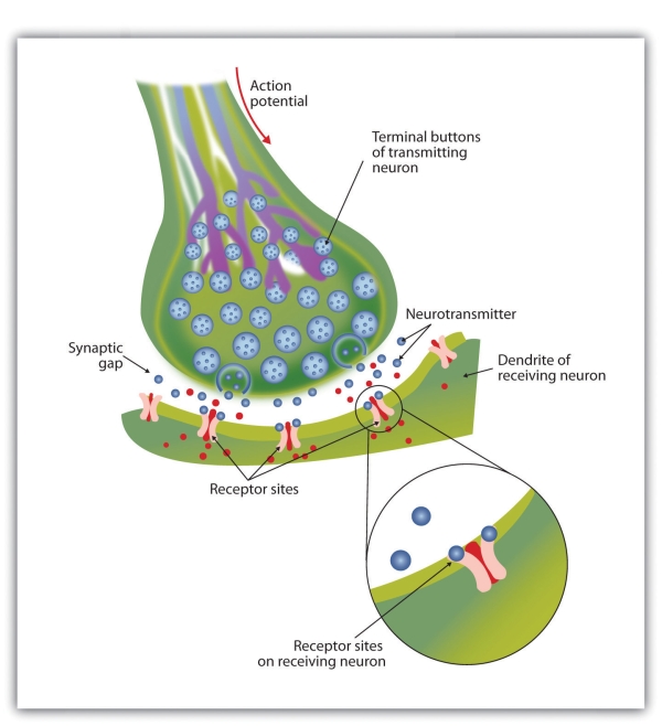
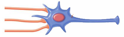
Not only do neural signals travel via electrical charges within the neuron, but they also travel via chemical transmission between the neurons. As we just learned, neurons are separated by junction areas known as synapses, areas where the terminal buttons at the end of the axon of one neuron nearly, but don’t quite, touch the dendrites of another. The synapses provide a remarkable function because they allow each axon to communicate with many dendrites in neighboring cells. Because a neuron may have synaptic connections with thousands of other neurons, the communication links among the neurons in the nervous system allow for a highly sophisticated communication system.
When the electrical impulse from the action potential reaches the end of the axon, it signals the terminal buttons to release neurotransmitters into the synapse. A neurotransmitter is a chemical that relays signals across the synapses between neurons. Neurotransmitters travel across the synaptic space between the terminal button of one neuron and the dendrites of other neurons, where they bind to the dendrites in the neighboring neurons.
Furthermore, different terminal buttons release different neurotransmitters, and different dendrites are particularly sensitive to different neurotransmitters. The dendrites admit the neurotransmitters only if they are the right shape to fit in the receptor sites on the receiving neuron. For this reason, the receptor sites and neurotransmitters are often compared to a lock and key.
“Introduction to Psychology: Unit 4, Module 5: Neurons Communicate Using Electrochemical Processes” by Open Learning Initiative. Retrieved from https://canvas.instructure.com/courses/773049/pages/stangor-3-dot-1-the-neuron-is-the-building-block-of-the-nervous-system. Licensed under CC BY-NC-SA.
When the nerve impulse reaches the terminal button, it triggers the release of neurotransmitters into the synapse. The neurotransmitters fit into receptors on the receiving dendrites in the manner of a lock and key.
When neurotransmitters are accepted by the receptors on the receiving neurons, their effect may be either excitatory (i.e., they make the cell more likely to fire) or inhibitory (i.e., they make the cell less likely to fire). Furthermore, if the receiving neuron is able to accept more than one neurotransmitter, it is influenced by the excitatory and inhibitory processes of each. If the excitatory effects of the neurotransmitters are greater than the inhibitory influences of the neurotransmitters, the neuron moves closer to its firing threshold, and if it reaches the threshold, the action potential and the process of transferring information through the neuron begins.
“Introduction to Psychology: Unit 4, Module 5: Neurons Communicate Using Electrochemical Processes” by Open Learning Initiative. Retrieved from https://canvas.instructure.com/courses/773049/pages/stangor-3-dot-1-the-neuron-is-the-building-block-of-the-nervous-system. Licensed under CC BY-NC-SA.
Postsynaptic Potentials
There are many “potentials” mentioned in neural communication – membrane potential, resting potential, action potential, threshold potential, and synaptic potential. To help keep them straight, note that the word “potential” essentially means “charge,” similar to the charge of the battery referencing its potential energy.
The many types of potentials (or “charges”):
- membrane potential – the charge of the plasma cell membrane of the neuron
- resting potential – the charge of the membrane while at rest; it happens that neurons have a net negative charge while at rest
- threshold potential – the charge of the membrane required to produce an action
- action potential – the charge of the membrane when having an action (sending an electrical signal)
- postsynaptic potential – the charge of the membrane due to the messages received across the synapse
To be more explicit, postsynaptic potentials are the result of neurotransmitters binding to the receptors on the dendrites of a neuron. This neuron is receiving information, hence the term “postsynaptic;” it is after the synapse. When these neurotransmitters bind, the neuron responds by opening or closing channels in its cell membrane, allowing various ions (charged particles) in and/or out. Recall the lock-and-key metaphor of such binding.
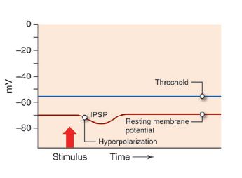
Because ions are, by definition, charged particles, they are either positive or negative. You can imagine, then, that an influx or efflux of such molecules will result in changes to the charge of the cell membrane, meaning that the membrane potential will either become more or less negative. Indeed, this concept is the underlying foundation of the two types of postsynaptic potentials. When the membrane becomes more negative, it has hyperpolarized (hyper = more, e.g. hyperactive). When the membrane becomes less negative (i.e. more positive), it has depolarized (de = less, e.g. decrease).
If the membrane becomes hyperpolarized when the ions move, an inhibitory postsynaptic potential (IPSP) is generated. Since it hyperpolarizes the membrane, the neuron is further away from the threshold potential.
IPSP: Opening of potassium- or chloride channels leads to hyperpolarization of the membrane. (Since the current is outward for potassium ions, and inward for chloride ions, opening of either of these two channels will cause the postsynaptic membrane to hyperpolarize.) A hyperpolarized membrane has moved farther from the threshold potential and has less probability of producing an action potential. Since an IPSP hyperpolarizes the membrane, it inhibits action potentials.
“Anatomy & Physiology: Unit 13, Module 54: Synaptic Potentials” by Open Learning Initiative. Retrieved from https://oli.cmu.edu/jcourse/workbook/activity/page?context=21af2e9d80020ca60036c9a001216067. Licensed under CC BY.
An excitatory postsynaptic potential (EPSP) occurs if the membrane is depolarized by the ion movement. An excitatory postsynaptic potential depolarizes the membrane, bringing it closer to the threshold potential.
EPSP: Opening of sodium- or calcium channels leads to depolarization of the membrane. If there is sufficient depolarization, the threshold potential is reached and an action potential will be produced in the postsynaptic membrane. Since an EPSP depolarizes the membrane, it facilitates action potentials.

“Anatomy & Physiology: Unit 13, Module 54: Synaptic Potentials” by Open Learning Initiative. Retrieved from https://oli.cmu.edu/jcourse/workbook/activity/page?context=21af2e9d80020ca60036c9a001216067. Licensed under CC BY.
And so, postsynaptic potentials are local potentials that develop in the postsynaptic cell’s membrane when neurotransmitters binding to receptors leads to the opening of ion channels.
Remember that a neuron synapses with many other neurons. So a postsynaptic neuron can receive signals from many presynaptic neurons simultaneously. Whether or not the postsynaptic cell has an action potential depends on the summation (the additive effect) of all the incoming signals. Each active synapse can result in a local potential (either an EPSP or an IPSP). The net effect of all the local potentials on the trigger zone determines whether or not there is an action potential in the postsynaptic cell.
There are two different ways that local potentials can sum to excite the postsynaptic cell to have an action potential. Temporal summation occurs when successive EPSPs at a single synapse occur in rapid succession. The successive potentials occur before the previous ones die out producing an increasing membrane depolarization.
“Anatomy & Physiology: Unit 14, Module 55: Synaptic Potentials” by Open Learning Initiative. Retrieved from https://oli.cmu.edu/jcourse/workbook/activity/page?context=21af2e9d80020ca60036c9a001216067. Licensed under CC-BY.
In other words, temporal summation occurs when one synapse stimulates the postsynaptic cell very quickly and the EPSPs produced in the postsynaptic cell piggyback on each other causing an increasing level of depolarization.
“Anatomy & Physiology: Unit 13, Module 54: Synaptic Potentials” by Open Learning Initiative. Retrieved from https://oli.cmu.edu/jcourse/workbook/activity/page?context=21af2e9d80020ca60036c9a001216067. Licensed under CC-BY.
Summation can also occur when multiple presynaptic neurons stimulate the postsynaptic neuron at the same time (spatial summation). Each individual synapse lets in a limited number of ions and alters the membrane potential a little. The collective effect of all the synapses allows enough ions in to reach the threshold potential and an action potential is triggered.
“Anatomy & Physiology: Unit 14, Module 55: Synaptic Potentials” by Open Learning Initiative. Retrieved from https://oli.cmu.edu/jcourse/workbook/activity/page?context=21af2e9d80020ca60036c9a001216067. Licensed under CC-BY.
In other words, spatial summation occurs when the collective effect of multiple synapses depolarizes the postsynaptic neuron to threshold resulting in an action potential.
The interplay between IPSPs and EPSPs is important. Whether an action potential is going to be produced depends not just on the summation of the EPSPs, but on the summation of EPSPs and IPSPs. The algebraic sum of all EPSPs and IPSPs has to be of sufficient amplitude to raise the membrane potential to the threshold for an action potential. Importantly, if the IPSPs prevail, then the post-synaptic cell will be “silent.” One can, therefore, visualize the process of summation as a “tug-of-war” between excitatory and inhibitory currents induced by the binding of neurotransmitters on excitatory or inhibitory postsynaptic receptors, respectively.
“Anatomy & Physiology: Unit 13, Module 54: Synaptic Potentials” by Open Learning Initiative. Retrieved from https://oli.cmu.edu/jcourse/workbook/activity/page?context=21af2e9d80020ca60036c9a001216067. Licensed under CC-BY.
Neural Communication – Video
CrashCourse. (2015, March 2). The nervous system, part 2 – Action! Potential!: Crash Course A&P #9.. Retrieved from https://www.youtube.com/watch?v=OZG8M_ldA1M&list=PL8dPuuaLjXtOAKed_MxxWBNaPno5h3Zs8&index=9. Standard YouTube License.
CrashCourse. (2015, March 10). The nervous system, Part 3 – Synapses!: Crash Course A&P #10. Retrieved from https://www.youtube.com/watch?v=VitFvNvRIIY&t=5s. Standard YouTube License.
Summary
The nervous system must receive input, process information, and respond to information; it follows, then, that the most basic units might have complementary abilities.
Neurons, just like other biological cells, have a cell membrane, which forms a boundary between intracellular fluid (cytoplasm) and extracellular fluid. Consider your own skin, which separates the interior of your body from the environment.
Because of this boundary, we can measure the cell’s membrane potential, which is the charge of its membrane, just like we can measure the temperature of your skin. Importantly, this membrane potential can change depending on its environment; similarly, you can become warmer or colder. The cell membrane has protein channels and pores, just like your skin, which allows it to interact with its environment.
Neurotransmitters bind to the receptors on a postsynaptic neuron, which results in the opening and/or closing of various channels, allowing different ions to flow in or out. These ions, being charged particles, alter the membrane potential – either increasing it or decreasing it. Without any stimulation, the cell is at rest; indeed, the membrane has a resting potential that is negative. In order to produce a signal itself, the membrane potential of the neuron must pass the threshold of excitation. In other words, your skin must become warm enough for you to be galvanized into action.
Different neurotransmitters have different effects on the membrane potential. If the membrane potential hyperpolarizes (decreases; becomes more negative), then the neuron is said to have experienced an inhibitory postsynaptic potential. The neuron is now further away from the threshold of excitation, and less likely to have an action potential; the neuron has been inhibited.
If the membrane potential depolarizes (increases; becomes less negative), then the neuron is said to have experienced an excitatory postsynaptic potential. The neuron is now closer to the threshold of excitation, and more likely to have an action potential; the neuron has been excited.
Remember that many neurotransmitters could be binding to their corresponding receptors, meaning that the neuron must summate (either temporally or spatially) the signals. Otherwise, we would have to receive all messages at exactly the same time and/or in exactly the same location, which would result in maladaptive signalling, just as it would for attempting to have a conversation with multiple people.
If this summation results in the neuron’s membrane potential exceeding the threshold of excitation, the neuron will have an action potential – the charge associated with sending a signal down the axon. This electrical signal will propagate down the axon, and result in the release of neurotransmitters (chemicals) from the terminal. As you can see, neural transmission is an electrochemical process.