Cells of the Nervous System
Introduction
Every behavior begins with biology. Our behaviors, as well as our thoughts and feelings, are produced by the actions of our brains, nerves, muscles, and glands. In this section we will begin our journey into the world of psychology by considering the biological makeup of the human being, including the most remarkable of human organs—the brain.
We will see that the body is controlled by an information highway known as the nervous system, a collection of hundreds of billions of specialized and interconnected cells through which messages are sent between the brain and the rest of the body. The nervous system consists of the central nervous system (CNS), made up of the brain and the spinal cord, and the peripheral nervous system (PNS), the neurons that link the CNS to our skin, muscles, and glands. And we will see that our behavior is also influenced in large part by the endocrine system, the chemical regulator of the body that consists of glands that secrete hormones.
The understanding of the biology underlying psychological processes is an important cornerstone of your new understanding of psychology. We will consider throughout the section how our biology influences important human behaviors, including our mental and physical health, our reactions to drugs, as well as our aggressive responses and our perceptions of other people. This section is particularly important for contemporary psychology because the ability to measure biological aspects of behavior, including the structure and function of the human brain, is progressing rapidly, and understanding the biological foundations of behavior is an increasingly important line of psychological study.
Stangor, C. (2017). Introduction to psychology. Boston, MA: Flatworld.
Cells of the Nervous System
Nervous system cells are considered the simplest functional unit of nervous tissue, and consist of two types: neurons and glia. Neurons are long-lived (most live for your entire life), electrically active cells that consume a lot of energy.
Just like your body and the cells that make up the body, these cells are constantly undergoing metabolism – taking in oxygen and glucose in order to sustain life processes. For this reason, it is utter nonsense to think that we only use 10% of our brains. If the remaining 90% of cells were not active (“in use”), they would die!
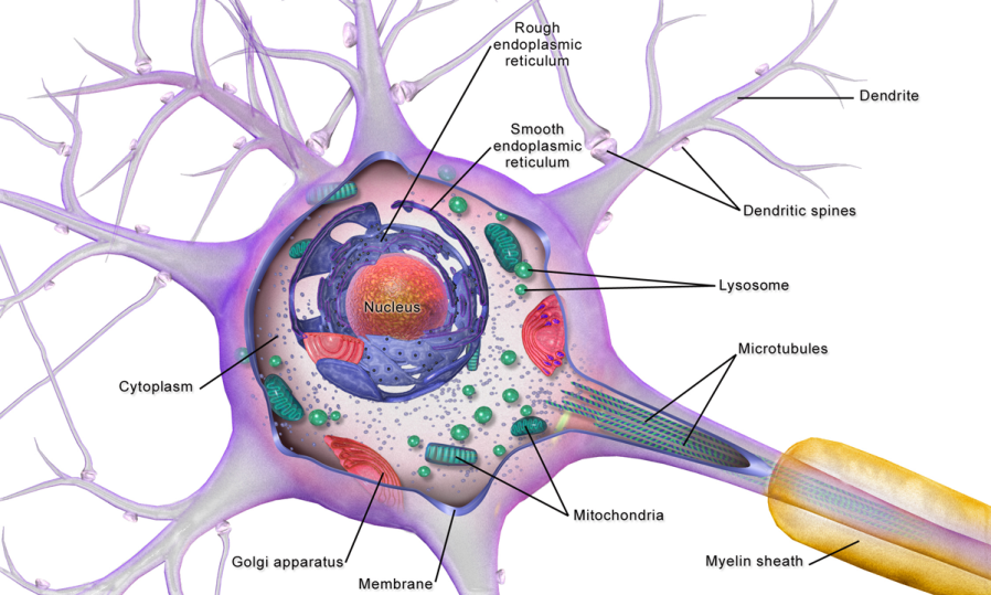
Neurons look like other biological cells: a plasma cell membrane, cytoplasm, and organelles, including nucleus, ribosomes, lysosomes, endoplasmic reticulum, mitochondria, etc. However, unlike other cells in the body, neurons have special appendages that allow them to communicate with each other. They have anatomically and functionally distinct regions for receiving, integrating and sending information from one part of the body to another.
Neurons are capable of responding to stimulation, conducting electrical signals, and secreting chemicals that allow them to communicate with other cells. They cannot usually regenerate if damaged since most neurons do not retain the ability to divide.
Glial cells exist in both the central and peripheral nervous systems and are the “glue” that hold these systems together by providing several supportive roles.
“Complete Neuron Cell Diagram” by LadyofHats. Retrieved from https://commons.wikimedia.org/wiki/File:Complete_neuron_cell_diagram_en.svg. Licensed under CC-BY.
“Anatomy & Physiology: Unit 13, Module 54: Neurons” by Open Learning Initiative. Retrieved from https://oli.cmu.edu/jcourse/workbook/activity/page?context=21af2e8280020ca60095320e001fa51f Licensed under CC BY.
Neurons
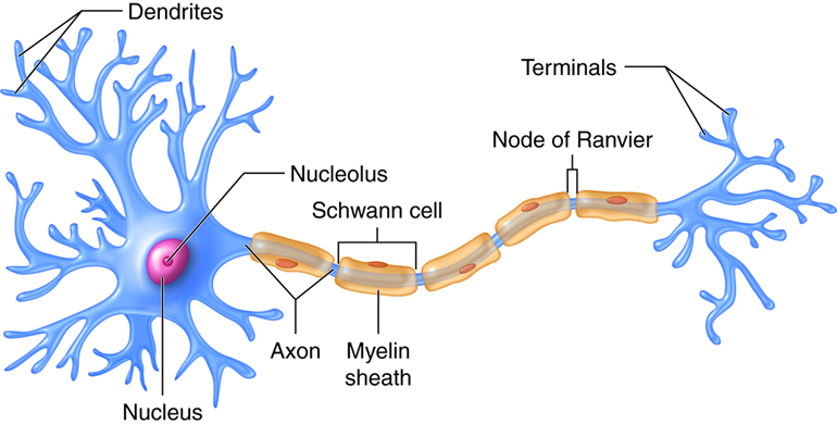
The nervous system is composed of more than 100 billion cells known as neurons. A neuron is a cell in the nervous system whose function is to receive and transmit information.
Neurons are made up of three major parts:
- a cell body, or soma, which contains the nucleus of the cell and keeps the cell alive
- a branching treelike fiber known as the dendrite, which collects information from other cells and sends the information to the soma
- a long, segmented fiber known as the axon, which transmits information away from the cell body toward other neurons or to the muscles and glands
Neurons are polarized with anatomically and chemically distinct regions. A typical neuron has two distinct processes or cytoplasmic extensions on either side of a soma (cell body).
On one side of the soma are short, tapering processes called dendrites (Greek, dendreon – tree). Most neurons have many, highly branched dendrites, although they may have as few as one. Dendrites receive information from other neurons and transfer it to the cell body. The greater the number of dendrites, the more information the neuron can collect to use during decision making.
The soma (cell body) is the region of the neuron that integrates all the incoming information from the dendrites. The cell body is somewhat spherical in shape and for humans, typically ranges in size from 5 – 100 microns in diameter. The soma contains the neuron’s nucleus and housekeeping organelles (e.g. mitochondria, lysosomes, Golgi complex, rough endoplasmic reticulum, etc.). The soma is the only site in a neuron that can synthesize proteins, neurotransmitters, or materials needed for cell maintenance and repair. The soma is similar to the dendrites in that it can also receive inputs from other neurons. The incoming information is coded as electrical signals that are integrated to determine what, if any, response is required.
If the incoming signal is large enough, it travels to the process on the other side of the soma called the axon. If the incoming signal is small, it dies out before it reaches the axon. The axon functions like a cable, relaying electrical signals away from the cell body towards other neurons or cells (e.g. muscles, glands). Axons are also called nerve fibers.
The axon has three regions. As it emerges from the cell body, the axon forms a structure called the axon hillock, a tapered region that contains the initial segment, or trigger zone, where propagating electrical signals called action potentials are initiated or generated. The next part of the axon is the longest, typically a single, thin (.5 – 3 microns), almost constant diameter process that extends to a target. Axons can be long, short, or in between.
For example, neurons that command the big toe muscles to contract have dendrites and cell bodies in the spinal cord and axons that leave the vertebral column to travel the length of the leg to the big toe. For a 7 foot tall basketball player, the axon that controls the big toe might be 3 feet long. On the other hand, short axons (a few microns long) are found on interneurons, neurons that have all processes within the central nervous system (CNS) and nearby targets.
The axon may travel to its target as a single fiber, but some axons form branches called collaterals, so that they can interact with not just one, but many target cells. The third region of the axon is found when it reaches its target. Here the axon branches extensively forming the synaptic terminals (terminal arborization). Axons branch out toward their ends, and at the tip of each branch is a terminal button, which contains vesicles filled with chemical messengers (neurotransmitters) that conduct the signal to the next cell.
“Anatomy & Physiology: Unit 13, Module 54: Neurons” by Open Learning Initiative. Retrieved from https://oli.cmu.edu/jcourse/workbook/activity/page?context=21af2e8280020ca60095320e001fa51fLicensed under CC BY.
Some neurons have hundreds or even thousands of dendrites, and these dendrites may themselves be branched to allow the cell to receive information from thousands of other cells. The axons are also specialized, and some, such as those that send messages from the spinal cord to the muscles in the hands or feet, may be very long—even up to several feet in length.
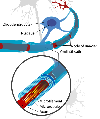
To improve the speed of their communication, and to keep their electrical charges from interacting with other neurons, axons are often surrounded by a myelin sheath. The myelin sheath is a layer of fatty tissue surrounding the axon of a neuron that both acts as an insulator and allows faster transmission of the electrical signal.
In a myelinated axon, the bare regions where the sheath is interrupted are called nodes of Ranvier. The myelinated segments between consecutive nodes of Ranvier are called internodes. The myelin sheath changes the appearance of axons as well as their electrical properties. Myelinated axons appear white when viewed by the naked eye in contrast to areas where neuronal cell bodies are concentrated, which appear gray.
It is this aesthetic difference that gives rise to the distinction of white matter (axons with myelin) versus gray matter (somas). Moreover, the gray matter is clustered on the outside of the brain, near the skull, forming cortex. The white matter is in the interior of the brain. This arrangement should be intuitive, as you consider the brain as a collection of processing units (somas) on the outside, with crossing insulated wires (axons) on the inside.
“Anatomy & Physiology: Unit 13, Module 54: Neurons” by Open Learning Initiative. Retrieved from https://oli.cmu.edu/jcourse/workbook/activity/page?context=21af2e8280020ca60095320e001fa51f Licensed under CC BY. “Introduction to Psychology: Chapter 3, Section 1: The Neuron Is the Building Block of the Nervous System” by the University of Minnesota Retrieved from http://open.lib.umn.edu/intropsyc/chapter/3-1-the-neuron-is-the-building-block-of-the-nervous-system/#stangor-ch03_s01_s01_n01 Licensed under CC BY-NC-SA.
Classification of Neurons: Structure
Neurons are classified according to two different characteristics: their morphology (anatomical or structural features) and their function.
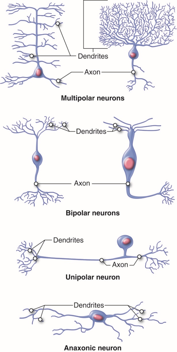
The structural classification of neurons is based on the number of processes that extend from the soma. There are four basic neuronal structures, though there are many subtle variations on each theme.
Bipolar neurons have a single dendrite extending from one side of the cell body and a single axon extending from the other side. Bipolar neurons are small cells, typically extending for less than 30 microns from dendrite to axon terminal. There are not many true bipolar cells in the body. A few examples are found in the special sense organs for vision and olfaction (smell).
Unipolar or pseudounipolar neurons have a single process that emanates from the cell body. The single process has dendrites on one end and the rest of the process is an axon. Most sensory neurons of the peripheral nervous system are unipolar neurons. The dendrites are located in the periphery, where stimuli are detected. The sensory information travels on the dendrite toward the soma (usually located ganglia just outside the CNS). The axon stretches into the CNS at the spinal cord.
Multipolar neurons have two or more dendrites on one side and a single axon on the other side of the soma. Multipolar neurons are the most common neurons in the CNS. One example are motor neurons which have dendrites and somas located in the spinal cord and axons that leave the CNS to innervate skeletal muscles.
Anaxonic neurons are small, stellate (star-shaped) cells with processes that all look alike with no apparent axon. Anaxonic neurons can be found in the central nervous system, the retina, and in the adrenal medulla. Their functions are not well understood.
“Anatomy & Physiology: Unit 13, Module 54: Neurons” by Open Learning Initiative. Retrieved from https://oli.cmu.edu/jcourse/workbook/activity/page?context=21af2e8280020ca60095320e001fa51fLicensed under CC BY.
“Anatomy & Physiology: Unit 13, Module 54: Neurons” by Open Learning Initiative. Retrieved from https://oli.cmu.edu/jcourse/workbook/activity/page?context=21af2e8280020ca60095320e001fa51f Licensed under CC BY.
Classification of Neurons: Function
Neurons are classified according to two different characteristics: their morphology (anatomical or structural features) and their function.
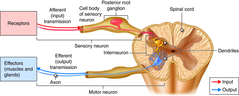
Functional classification of neurons is based on the direction of information flow along axons relative to the central nervous system (CNS). Based on this criterion, there are three types of neurons: sensory neurons, interneurons, and motor neurons.
Sensory (afferent) neurons are specialized for detection of sensory information (e.g. light, pressure, vibration, temperature, chemicals, etc.). They transduce physical and chemical stimuli into electrical signals and transfer this information from the periphery towards the central nervous system for processing. In many cases, sensory neurons have their dendrites, soma and a part of their axon residing outside the CNS with axon terminals forming connections (synapses) with other neurons within the CNS.
Interneurons (association neurons) are located entirely within the central nervous system (with the dendrites, soma and axons of the cell all residing within the CNS). Interneurons are also referred to as association neurons, in part because they are sandwiched between sensory and motor neurons where they integrate and distribute sensory information and coordinate motor output. Interneurons account for 90% of all neurons of the CNS and therefore are the most numerous neurons in the body. Almost all interneurons are multipolar.
Motor (efferent) neurons carry impulses or motor commands away from the central nervous system to effectors/target organs (e.g. muscles and glands). Most motor neurons have dendrites and cell bodies in the CNS and axons that exit the CNS to form peripheral nerves that travel to effectors (targets).
“Anatomy & Physiology: Unit 13, Module 54: Neurons” by Open Learning Initiative. Retrieved from https://oli.cmu.edu/jcourse/workbook/activity/page?context=21af2e8280020ca60095320e001fa51fLicensed under CC BY.
“Anatomy & Physiology: Unit 13, Module 54: Neurons” by Open Learning Initiative. Retrieved from https://oli.cmu.edu/jcourse/workbook/activity/page?context=21af2e8280020ca60095320e001fa51f Licensed under CC BY.
Glial Cells
Most neurons are surrounded by glial cells (neuroglia), the other cell type found in the nervous tissue. Glial cells (or glia, for short) are incredibly important, but often receive less attention. Glial cells are the supportive cells of the nervous system, and are 10 times more numerous than neurons!
The most well defined role for neuroglia is to provide structure to the delicate nervous tissue. They fill the space between neurons, serving as mortar or “glue” and thus hold nervous tissue together. (Indeed, glia means “glue.”)
Unlike neurons, glial cells retain the ability to divide throughout one’s lifetime. When neurons are injured, neuroglia are stimulated to divide and form glial scars. Glial cells have different shapes and sizes and their processes are indistinguishable in contrast to the distinct axon and dendrites found in neurons.
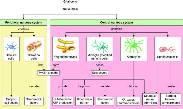
There are six types of glial cells, four types are found in the CNS and two types in the PNS. The four types of glia found in the central nervous system (CNS) are: astrocytes; oligodendrocytes; microglia, and ependymal cells. The two types of glia found only in the peripheral nervous system (PNS) are satellite cells and Schwann cells.
“Anatomy & Physiology: Unit 13, Module 54: Neurons” by Open Learning Initiative. Retrieved from https://oli.cmu.edu/jcourse/workbook/activity/page?context=21af2e8280020ca60095320e001fa51fLicensed under CC BY.
“Anatomy & Physiology: Unit 13, Module 54: Neurons” by Open Learning Initiative. Retrieved from https://oli.cmu.edu/jcourse/workbook/activity/page?context=21af2e8280020ca60095320e001fa51f Licensed under CC BY.
Glial Cells in the Central Nervous System (CNS)
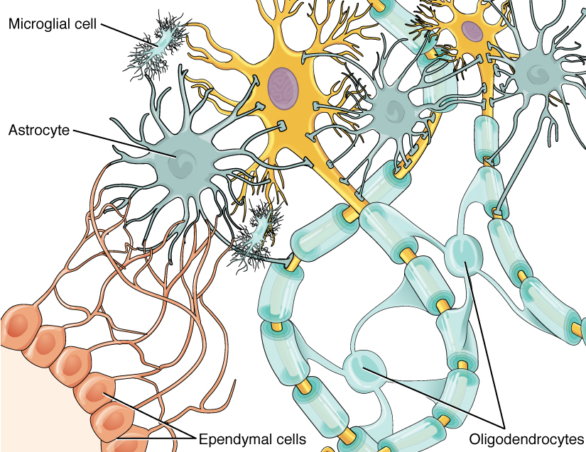
Astrocytes are star-shaped neuroglia and are the most numerous cells in the central nervous system. They make up half of all cells in the brain. Astrocytes provide a structurally supportive framework for neurons with their processes wrapping most non-synaptic regions of neurons in gray matter and covering the entire outer surface of the brain to form the glial – pia (connective tissue meninx) interface.
Astrocytes help form the protective blood-brain barrier by encircling CNS capillary endothelial cells and stimulating the cells to form tight-junctions. They help to maintain the concentration of chemicals in the extracellular space and remove excess signalling molecules. Astrocytes also react to neural tissue damage by forming scar tissue in the damaged space.
Oligodendrocytes are glial cells of the CNS that wrap and insulate axons and give the CNS white matter its characteristic glossy, white appearance. Oligodendrocytes have a large soma with up to 15 processes. The processes reach out to axons of nearby neurons and wrap around them (like wrapping tape around a pencil) forming a high resistance sheath called myelin.
Myelin insulates a small region of the axon (prevents ions from leaking out into the extracellular fluid), which facilitates signal propagation down the axon towards the synaptic terminal. A single oligodendrocyte’s processes will wrap axons of numerous different neurons. Processes from many different oligodendrocytes contribute to the myelin sheath of a single neuron’s axon.
Microglia are small highly mobile, phagocytic neuroglia that protect nervous tissue from pathogen infection, remove debris and waste, and may play a role in remodeling of the synapse that occurs during development and with learning. About 10-15% of CNS glial cells are microglia. Microglia are derived from monocytes and thus are more closely related to white blood cells than to the other glial cells.
Since cells of the immune system cannot penetrate the blood brain barrier, microglia serve as brain macrophages, destroying foreign invaders, promoting inflammation and destroying cancer cells and cells infected with virus. Clusters of microglia in nervous tissue provide pathologists with evidence of recent injury.
Ependymal cells are cuboidal-shaped glial cells that are joined together to form a continuous sheet lining the fluid-filled ventricles and central canal of the brain and spinal cord. Ependymal cells produce and secrete cerebrospinal fluid (CSF), the fluid that bathes the tissues of the CNS. The basal side of the cell has rootlets that anchor the cells to the underlying tissue. The apical surface is marked by cilia, which helps circulate the CSF.
“Neuron with Oligodendrocyte and Myelin Sheath” by Andrew C. Retrieved from https://commons.wikimedia.org/w/index.php?search=neuron+histology&title=Special:Search&profile=images&fulltext=1&searchToken=dcpxvxldsu7s7f26cw7rkhkn2#/media/File:Neuron_with_oligodendrocyte_and_myelin_sheath.svg. Licensed under CC-BY.
“Anatomy & Physiology: Unit 13, Module 54: Neurons” by Open Learning Initiative. Retrieved from https://oli.cmu.edu/jcourse/workbook/activity/page?context=21af2e8280020ca60095320e001fa51f Licensed under CC BY.
Glial Cells in the Peripheral Nervous System
The remaining two glial cells, Schwann cells and satellite cells, are found solely in the peripheral nervous system.
Schwann cells are analogous in function to oligodendrocytes (found in the CNS). They insulate the axons of peripheral nerves in one of two ways. A Schwann cell can wind its way round and round the axon (up to 100 times), while squeezing its cytoplasm out of the way (much like a toothpaste tube could be wrapped around a pencil), forming a myelin sheath. Like myelinating a single fiber in the CNS, which requires many oligodendrocytes, a complete myelin sheath in the PNS requires many Schwann cells. Schwann cells can also envelop PNS axons without forming a myelin sheath. Instead of wrapping a single axon many times, the Schwann cell forms an envelope around a bundle of unmyelinated axons.
Additionally, Schwann cells can also assist in the regeneration of a damaged peripheral nerve. If a peripheral nerve is damaged, it may regenerate if its soma is undamaged and the neurilemma (the plasma membrane of the Schwann cell) enveloping it is intact.
Satellite cells are found surrounding neural somas in peripheral ganglia (collections of cell bodies located outside the CNS). Satellite cells resemble CNS astrocytes and are thought to have similar functions, providing structural support and regulating the chemical environment.
Neuronal axons in the CNS and PNS can be devoid of a glial cell wrap (unmyelinated) or they can be discontinuously wrapped by glial cells along their entire length (myelinated).

“Glia” by BruceBlaus. Retrieved from https://commons.wikimedia.org/wiki/File:Glia.png. Licensed under CC-BY-SA-4.0.
“Glial Cells of the CNS” by OpenStax. Retrieved from https://commons.wikimedia.org/wiki/File:1209_Glial_Cells_of_the_CNS-02.jpg. Licensed under CC-BY-4.0.
“Anatomy & Physiology: Unit 13, Module 54: Neurons” by Open Learning Initiative. Retrieved from https://oli.cmu.edu/jcourse/workbook/activity/page?context=21af2e8280020ca60095320e001fa51f Licensed under CC BY.
Cells of the Nervous System
CrashCourse. (2015, February 23). The nervous system, Part 1: Crash Course A&P #8.Retrieved from https://www.youtube.com/watch?v=qPix_X-9t7E. Standard YouTube License.
Summary
Neurons are cells in the central nervous system, with specialized appendages to allow for electrochemical intercellular communication. These appendages project from the soma: the dendrites are branching extensions that receive information from other neurons, whereas the axon is a fiber that conducts an electrical signal to its terminals. This axon, just like an electrical wire, is insulated in myelin, with gaps called nodes of Ranvier, which increase the rate of signal transmission via saltatory conduction.
However, neurons are not the only type of cell in the nervous system. Some glial cells are in the central nervous system (CNS), whereas others are only found in the peripheral nervous system (PNS).
Found in the CNS are astrocytes, oligodendrocytes, microglia, and ependymal cells. Astrocytes provide structural support and regulate the chemical environment, whereas oligodendrocytes form myelin around the axons of neurons. Microglia are the janitors of the brain, clearing out waste products, whereas ependymal cells form the boundaries of ventricles, the spaces in the brain which contain cerebrospinal fluid (CSF) for cushioning.
Found in the PNS are Schwann cells, which form myelin similarly to oligodendrocytes, and satellite cells, which provide structural support and regulate the chemical environment similar to astrocytes.