Structures of the Brain
Introduction
If you were someone who understood brain anatomy and were to look at the brain of an animal that you had never seen before, you would nevertheless be able to deduce the likely capacities of the animal. This ability is because the brains of all animals are very similar in overall form.
In each animal, the brain is layered, and the basic structure of the brain is similar. Indeed, anatomists often state that the structure of the brain is conserved across species, which affords scientists the ability to make cross-species comparisons when studying processes.
Systems and tissues of the brain can be discussed in a variety of ways, listed below:
- early development
- evolutionary age (“old” to “new”)
- structure and function
This material will discuss various ways that one can categorize and discuss portions of the brain. Importantly, depending on the method of categorization, the subcategories will differ. For example, if one were to divide the population by religious practices, the subcategories might include: Christianity, Hinduism, Islam, Judaism, etc. If one were to divide the population by ethnicity, the subcategories might include: African-American, Caucasian, Asian-American, etc.
Critically, an individual person could be “labelled” in a variety of ways, depending on the category chosen. We can draw a parallel to the brain. There are many brain regions that could be discussed in a variety of ways, depending on the method of categorization.
“Introduction to Psychology: Chapter 3, Section 2: Our Brains Control Our Thoughts, Feelings, and Behavior” by the University of Minnesota Retrieved from http://open.lib.umn.edu/intropsyc/chapter/3-2-our-brains-control-our-thoughts-feelings-and-behavior/ Licensed under CC BY-NC-SA.
Parts of the Brain
Early Development of Systems
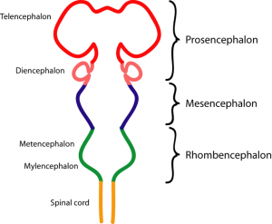
From the neural tube, the brain develops three systems of tissue: forebrain, midbrain, and hindbrain. These latter two regions, the midbrain and the hindbrain, are often grouped together and called the brainstem.
Notably, during early development of the fetus, these three regions are discussed in more complicated terms. The forebrain, or prosencephalon, consists of two subregions: the telencephalon and the diencephalon. The midbrain, or mesencephalon, does not have any subregions during early development. The hindbrain, or rhombencephalon, consists of two subregions: the metencephalon and the myelencephalon.
Now, these words are intimidating, but realize that the word “encephalon” simply means “brain.” Indeed, encephalitis is simply an –itis (disease or inflammation) of the encephalon (brain). So, each of these more complicated terms are essentially just specifying a portion of the brain.
The telencephalon becomes the cerebrum of the brain, or the “bulk” of it, which contains the wrinkly portion on the outside – “cortex”—and the innermost portions thereof.
“Embryonic Brain” by MrArifnajafov. Retrieved from https://commons.wikimedia.org/wiki/File:EmbryonicBrain_az.png. Licensed under CC0-1.0.
From “Old” to “New”
Old Brain
The innermost structures of the brain—the parts nearest the spinal cord—are the “oldest” part of the brain, evolutionarily-speaking, and these areas carry out the same functions they did for our distant ancestors. The “old brain” regulates basic survival functions, such as breathing, moving, resting, and feeding, and creates our experiences of emotion.
The “old brain” consists of the following regions:
- brainstem
- medulla
- pons
- reticular formation
- thalamus
- cerebellum
- limbic system (amygdala, hypothalamus, hippocampus)
“Introduction to Psychology: Chapter 3, Section 2: Our Brains Control Our Thoughts, Feelings, and Behavior” by the University of Minnesota.Retrieved from http://open.lib.umn.edu/intropsyc/chapter/3-2-our-brains-control-our-thoughts-feelings-and-behavior/Licensed under CC BY-NC-SA.
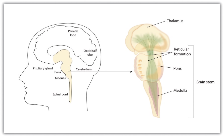
The brainstem is the oldest and innermost region of the brain. It’s designed to control the most basic functions of life, including breathing, attention, and motor responses. The brainstem begins where the spinal cord enters the skull and forms the medulla, the area of the brainstem that controls heart rate and breathing. In many cases, the medulla alone is sufficient to maintain life—animals that have the remainder of their brains above the medulla severed are still able to eat, breathe, and even move. The spherical shape above the medulla is the pons, a structure in the brain stem that helps control the movements of the body, playing a particularly important role in balance and walking.
Running through the medulla and the pons is a long, narrow network of neurons known as the reticular formation. The job of the reticular formation is to filter out some of the stimuli that are coming into the brain from the spinal cord and to relay the remainder of the signals to other areas of the brain. The reticular formation also plays important roles in walking, eating, sexual activity, and sleeping. When electrical stimulation is applied to the reticular formation of an animal, it immediately becomes fully awake, and when the reticular formation is severed from the higher brain regions, the animal falls into a deep coma.
Above the brain stem are other parts of the old brain that also are involved in the processing of behavior and emotions. The thalamus is the egg-shaped structure above the brain stem that applies more filtering to the sensory information that is coming up from the spinal cord and through the reticular formation, and it relays some of these remaining signals to the higher brain levels (Guillery & Sherman, 2002). The thalamus also receives some of the higher brain’s replies, forwarding them to the medulla and the cerebellum. The thalamus is also important in sleep because it shuts off incoming signals from the senses, allowing us to rest.
The cerebellum (literally, “little brain”) consists of two wrinkled ovals behind the brain stem. It functions to coordinate voluntary movement. People who have damage to the cerebellum have difficulty walking, keeping their balance, and holding their hands steady. Consuming alcohol influences the cerebellum, which is why people who are drunk have more difficulty walking in a straight line. Also, the cerebellum contributes to emotional responses, helps us discriminate between different sounds and textures, and is important in learning (Bower & Parsons, 2003).
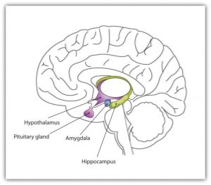
Whereas the primary function of the brain stem is to regulate the most basic aspects of life, including motor functions, the limbic system is largely responsible for memory and emotions, including our responses to reward and punishment. The limbic system is a brain area, located between the brain stem and the two cerebral hemispheres, that governs emotion and memory. It includes the amygdala, the hypothalamus, and the hippocampus.
“Introduction to Psychology: Chapter 3, Section 2: Our Brains Control Our Thoughts, Feelings, and Behavior” by the University of Minnesota.Retrieved from http://open.lib.umn.edu/intropsyc/chapter/3-2-our-brains-control-our-thoughts-feelings-and-behavior/Licensed under CC BY-NC-SA.
The amygdala consists of two “almond-shaped” clusters (amygdala comes from the Latin word for “almond”) and is primarily responsible for regulating our perceptions of, and reactions to, aggression and fear. The amygdala has connections to other bodily systems related to fear, including the sympathetic nervous system (which we will see later is important in fear responses), facial responses (which perceive and express emotions), the processing of smells, and the release of neurotransmitters related to stress and aggression (Best, 2009).
In one early study, Klüver and Bucy (1939) damaged the amygdala of an aggressive rhesus monkey. They found that the once angry animal immediately became passive and no longer responded to fearful situations with aggressive behavior. Electrical stimulation of the amygdala in other animals also influences aggression. In addition to helping us experience fear, the amygdala also helps us learn from situations that create fear. When we experience events that are dangerous, the amygdala stimulates the brain to remember the details of the situation so that we learn to avoid it in the future (Sigurdsson, Doyère, Cain, & LeDoux, 2007).
Located just under the thalamus (hence its name), the hypothalamus is a brain structure that contains a number of small areas that perform a variety of functions, including the important role of linking the nervous system to the endocrine system via the pituitary gland. Through its many interactions with other parts of the brain, the hypothalamus helps regulate body temperature, hunger, thirst, and sex, and responds to the satisfaction of these needs by creating feelings of pleasure. Olds and Milner (1954) discovered these reward centers accidentally after they had momentarily stimulated the hypothalamus of a rat.
The researchers noticed that after being stimulated, the rat continued to move to the exact spot in its cage where the stimulation had occurred, as if it were trying to re-create the circumstances surrounding its original experience. Upon further research into these reward centers, Olds (1958) discovered that animals would do almost anything to re-create enjoyable stimulation, including crossing a painful electrified grid to receive it. In one experiment a rat was given the opportunity to electrically stimulate its own hypothalamus by pressing a pedal. The rat enjoyed the experience so much that it pressed the pedal more than 7,000 times per hour until it collapsed from sheer exhaustion.
The hippocampus consists of two “horns” that curve back from the amygdala. The hippocampus is important in storing information in long-term memory. If the hippocampus is damaged, a person cannot build new memories, living instead in a strange world where everything he or she experiences just fades away, even while older memories from the time before the damage are untouched.
“Introduction to Psychology: Chapter 3, Section 2: Our Brains Control Our Thoughts, Feelings, and Behavior” by the University of Minnesota. Retrieved from http://open.lib.umn.edu/intropsyc/chapter/3-2-our-brains-control-our-thoughts-feelings-and-behavior/ Licensed under CC BY-NC-SA.
New Brain
Mammals, including humans, have developed further brain layers that provide more advanced functions—for instance, better memory, more sophisticated social interactions, and the ability to experience emotions. Humans have a very large and highly developed outer layer known as the cerebral cortex (the “wrinkly stuff”), which makes us particularly adept at these high-level processes.
All animals have adapted to their environments by developing abilities that help them survive. Some animals have hard shells, others run extremely fast, and some have acute hearing. Human beings do not have any of these particular characteristics, but we do have one big advantage over other animals—we are very, very smart.
You might think that we should be able to determine the intelligence of an animal by looking at the ratio of the animal’s brain weight to the weight of its entire body. But this does not really work. The elephant’s brain is one thousandth of its weight, but the whale’s brain is only one ten-thousandth of its body weight. On the other hand, although the human brain is one 60th of its body weight, the mouse’s brain represents one fortieth of its body weight. Despite these comparisons, elephants do not seem 10 times smarter than whales, and humans definitely seem smarter than mice.
The key to the advanced intelligence of humans is not found in the size of our brains. What sets humans apart from other animals is our larger cerebral cortex—the outer bark-like layer of our brain that allows us to so successfully use language, acquire complex skills, create tools, and live in social groups (Gibson, 2002). In humans, the cerebral cortex is wrinkled and folded, rather than smooth as it is in most other animals. This folding creates a much greater surface area and size, and allows increased capacities for learning, remembering, and thinking. The folding of the cerebral cortex is referred to as corticalization.
Although the cortex is only about one tenth of an inch thick, it makes up more than 80% of the brain’s weight. The cortex contains about 20 billion nerve cells and 300 trillion synaptic connections (de Courten-Myers, 1999). Supporting all these neurons are billions more glial cells (glia), cells that surround and link to the neurons, protecting them, providing them with nutrients, and absorbing unused neurotransmitters. The glia come in different forms and have different functions. For instance, the myelin sheath surrounding the axon of many neurons is a type of glial cell. The glia are essential partners of neurons, without which the neurons could not survive or function (Miller, 2005).
“Introduction to Psychology: Chapter 3, Section 2: Our Brains Control Our Thoughts, Feelings, and Behavior” by the University of Minnesota. Retrieved from http://open.lib.umn.edu/intropsyc/chapter/3-2-our-brains-control-our-thoughts-feelings-and-behavior/ Licensed under CC BY-NC-SA.
Structure and Function of the Cortex
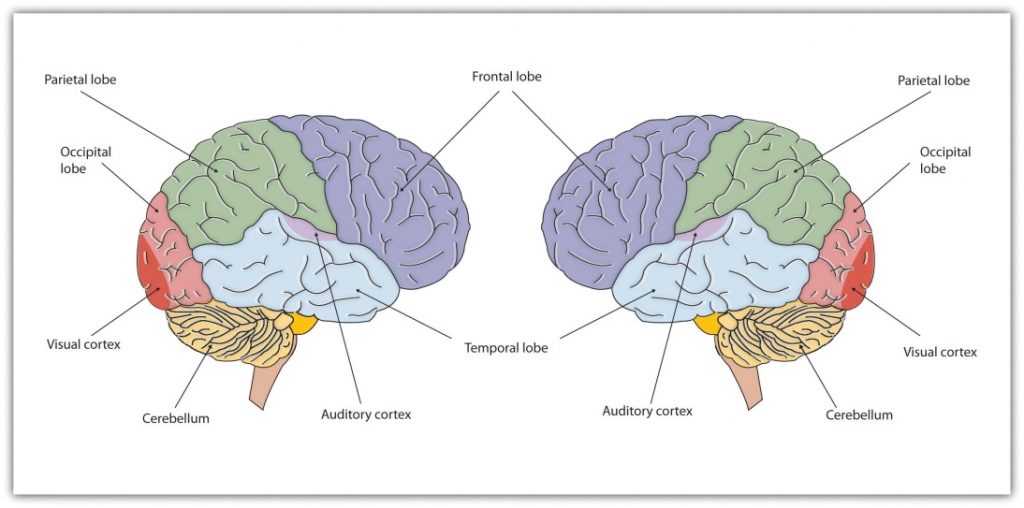
The cerebral cortex is divided into two hemispheres, and each hemisphere is divided into four lobes, each separated by folds known as fissures. If we look at the cortex starting at the front of the brain and moving over the top, we see the frontal lobe (behind the forehead), which is responsible primarily for thinking, planning, memory, and judgment. Following the frontal lobe is the parietal lobe, which extends from the middle to the back of the skull and which is responsible primarily for processing information about touch. Then comes the occipital lobe, at the very back of the skull, which processes visual information. Finally, in front of the occipital lobe (pretty much between the ears) is the temporal lobe, responsible primarily for hearing and language.
When the German physicists Gustav Fritsch and Eduard Hitzig (1870/2009) applied mild electric stimulation to different parts of a dog’s cortex, they discovered that they could make different parts of the dog’s body move. Furthermore, they discovered an important and unexpected principle of brain activity. They found that stimulating the right side of the brain produced movement in the left side of the dog’s body, and vice versa. This finding follows from a general principle about how the brain is structured, called contralateral control. The brain is wired such that in most cases the left hemisphere receives sensations from and controls the right side of the body, and vice versa.
“Introduction to Psychology: Chapter 3, Section 2: Our Brains Control Our Thoughts, Feelings, and Behavior” by the University of MinnesotaRetrieved from http://open.lib.umn.edu/intropsyc/chapter/3-2-our-brains-control-our-thoughts-feelings-and-behavior/Licensed under CC BY-NC-SA.
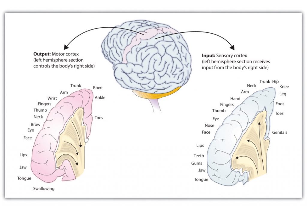
Fritsch and Hitzig also found that the movement that followed the brain stimulation only occurred when they stimulated a specific arch-shaped region that runs across the top of the brain from ear to ear, just at the front of the parietal lobe. Fritsch and Hitzig had discovered the motor cortex, the part of the cortex that controls and executes movements of the body by sending signals to the cerebellum and the spinal cord.
More recent research has mapped the motor cortex even more fully by providing mild electronic stimulation to different areas of the motor cortex in fully conscious patients while observing their bodily responses (because the brain has no sensory receptors, these patients feel no pain). This research has revealed that the motor cortex is specialized for providing control over the body, in the sense that the parts of the body that require more precise and finer movements, such as the face and the hands, also are allotted the greatest amount of cortical space.
Just as the motor cortex sends out messages to the specific parts of the body, the somatosensory cortex, an area just behind and parallel to the motor cortex at the back of the frontal lobe, receives information from the skin’s sensory receptors and the movements of different body parts. Again, the more sensitive the body region, the more area is dedicated to it in the sensory cortex. Our sensitive lips, for example, occupy a large area in the sensory cortex, as do our fingers and genitals.
The portion of the sensory and motor cortex devoted to receiving messages that control specific regions of the body is determined by the amount of fine movement that area is capable of performing. Thus, the hand and fingers have as much area in the cerebral cortex as does the entire trunk of the body.
Other areas of the cortex process other types of sensory information. The visual cortex is the area located in the occipital lobe (at the very back of the brain) that processes visual information. If you were stimulated in the visual cortex, you would see flashes of light or color, and perhaps you remember having had the experience of “seeing stars” when you were hit in, or fell on, the back of your head. The temporal lobe, located on the lower side of each hemisphere, contains the auditory cortex, which is responsible for hearing and language. The temporal lobe also processes some visual information, providing us with the ability to name the objects around us (Martin, 2007).
The motor and sensory areas of the cortex account for a relatively small part of the total cortex. The remainder of the cortex is made up of association areas in which sensory and motor information is combined and associated with our stored knowledge. These association areas are the places in the brain that are responsible for most of the things that make human beings seem human. The association areas are involved in higher mental functions, such as learning, thinking, planning, judging, moral reflecting, figuring, and spatial reasoning.
“Introduction to Psychology: Chapter 3, Section 2: Our Brains Control Our Thoughts, Feelings, and Behavior” by the University of Minnesota.Retrieved from http://open.lib.umn.edu/intropsyc/chapter/3-2-our-brains-control-our-thoughts-feelings-and-behavior/Licensed under CC BY-NC-SA.
“Introduction to Psychology: Chapter 3, Section 2: Our Brains Control Our Thoughts, Feelings, and Behavior” by the University of Minnesota. Retrieved from http://open.lib.umn.edu/intropsyc/chapter/3-2-our-brains-control-our-thoughts-feelings-and-behavior/ Licensed under CC BY-NC-SA.
Neuroplasticity
The control of some specific bodily functions, such as movement, vision, and hearing, is performed in specified areas of the cortex, and if these areas are damaged, the individual will likely lose the ability to perform the corresponding function. For instance, if an infant suffers damage to facial recognition areas in the temporal lobe, it is likely that he or she will never be able to recognize faces (Farah, Rabinowitz, Quinn, & Liu, 2000).
On the other hand, the brain is not divided up in an entirely rigid way. The brain’s neurons have a remarkable capacity to reorganize and extend themselves to carry out particular functions in response to the needs of the organism, and to repair damage. As a result, the brain constantly creates new neural communication routes and rewires existing ones. Neuroplasticity refers to the brain’s ability to change its structure and function in response to experience or damage. Neuroplasticity enables us to learn and remember new things and adjust to new experiences.
Our brains are the most “plastic” when we are young children, as it is during this time that we learn the most about our environment. On the other hand, neuroplasticity continues to be observed even in adults (Kolb & Fantie, 1989). The principles of neuroplasticity help us understand how our brains develop to reflect our experiences. For instance, accomplished musicians have a larger auditory cortex compared with the general population (Bengtsson et al., 2005) and also require less neural activity to move their fingers over the keys than do novices (Münte, Altenmüller, & Jäncke, 2002). These observations reflect the changes in the brain that follow our experiences.
Plasticity is also observed when there is damage to the brain or to parts of the body that are represented in the motor and sensory cortexes. When a tumor in the left hemisphere of the brain impairs language, the right hemisphere will begin to compensate to help the person recover the ability to speak (Thiel et al., 2006). And if a person loses a finger, the area of the sensory cortex that previously received information from the missing finger will begin to receive input from adjacent fingers, causing the remaining digits to become more sensitive to touch (Fox, 1984).
Although neurons cannot repair or regenerate themselves as skin or blood vessels can, new evidence suggests that the brain can engage in neurogenesis, the forming of new neurons (Van Praag, Zhao, Gage, & Gazzaniga, 2004). These new neurons originate deep in the brain and may then migrate to other brain areas where they form new connections with other neurons (Gould, 2007). This observation leaves open the possibility that someday scientists might be able to “rebuild” damaged brains by creating drugs that help grow neurons.
“Introduction to Psychology: Chapter 3, Section 2: Our Brains Control Our Thoughts, Feelings, and Behavior” by the University of Minnesota. Retrieved from http://open.lib.umn.edu/intropsyc/chapter/3-2-our-brains-control-our-thoughts-feelings-and-behavior/. Licensed under CC BY-NC-SA.”Introduction to Psychology: Unit 4, Module 6: The Cerebral Cortex” by Open Learning Initiative. Retrieved from https://oli.cmu.edu/jcourse/workbook/activity/page?context=df3e70800a0001dc71b2b133dc0cae8e. Licensed under CC BY-NC-SA.
Parts of the Brain – Video
CrashCourse. (2014, February 24). Meet your master: Getting to know your brain – Crash Course Psychology #4. Retrieved from https://www.youtube.com/watch?v=vHrmiy4W9C0. Standard YouTube License.
Physiology with Christian. (2017, October 25). How the human brain is different from other animals. Retrieved from https://www.youtube.com/watch?v=PydsQSNrcSo. Standard YouTube License.
Summary
The structure of animal brains is remarkably similar, showing conservation across species. It is often difficult to teach and understand the different divisions and parts of the brain due to a misunderstood or overlapping categorization. In other words, although there are many terms that can be used when discussing the brain, they belong to different categories.
The earlier material discussed the brain in the following ways:
- early development
- evolutionary age (“old” to “new”)
- structure and function
Regarding early development, the brain can be labelled as such:
- (forebrain) prosencephalon: telencephalon, diencephalon
- (midbrain) mesencephalon
- (hindbrain) rhombencephalon: metencephalon, myelencephalon
Regarding evolutionary age (“old” to “new”), the brain can be labelled as such:
- brainstem
- medulla
- pons
- reticular formation
- thalamus
- cerebellum
- limbic system (amygdala, hypothalamus, hippocampus)
- cortex
Regarding structure and function, the brain’s cerebral cortex is divided into two hemispheres (left and right), each of which has four lobes:
- frontal lobe – executive function, personality, decision-making
- parietal lobe – motor cortex, somatosensory cortex
- temporal lobe – memory, auditory cortex
- occipital lobe – visual cortex
Assessment Questions: “Anatomy and Physiology: Chapter 13, Section 1: The Embryologic Perspective” by OpenStax. Retrieved from https://opentextbc.ca/anatomyandphysiology/chapter/13-1-the-embryologic-perspective/. Licensed under CC BY.