Microscopy
Microscopy
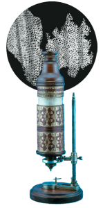
You may already know that the invention of the microscope led to the discovery of cells – but did you know that it was the visual appearance of dead cork cells through the microscope which led to their name (“cells”)? In 1665, an English scientist named Robert Hooke first observed tiny shavings of cork through his newly invented device, the compound microscope.
His drawing showed a honeycomb-like network of tiny little boxes all connected together like small rooms. Based upon this observation, Robert Hooke decreed these structures to be ‘cellulae’, which is Latin for ‘little storage rooms’. As it was known that the structures observed were from dried, nonliving bark, Hooke later wrote that in living trees and other plants, ‘…these cells are filled with juices.’
Subsequent improvements of the microscope were made by researchers like Anton Von Leeuwenhoek, who was the first to discover mobile, microscopic life forms present in pond water such as those seen in the following video:
As noted in your text (Science in Action: The Search for the Cell), prior to these observations, many smaller life forms, such as fleas, aphids, and weevils, were thought to spontaneously appear out of simple sand or dust.

Microscope Types
All microscopes are tools which are used to allow visualization of structures that are simply too small to see with the naked eye. There are three main types of microscopes which we will discuss now:
- Light Microscope
- Transmission Electron Microscope
- Scanning Electron Microscope
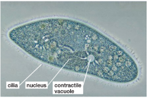
1) Light Microscope
Compound light microscopes use a series of glass lenses in series to bend and focus light to magnify images. The key advantage of the light microscope is that it can be used to observe living material. Light microscopes can achieve a level of magnification up to 1000X, enabling observation of structures as small as 20 nanometers (1 nanometer = 1 billionth of a meter).
2) Transmission Electron Microscope (TEM)
In contrast to light microscopes, electron microscopes use a powerful electron beam focused by magnetic fields to view structures. Another fundamental difference with electron microscopes is that they can only be used to view dead material that has been processed and coated with metals to reflect the electrons in the electron beam (similar to the way an x-ray machine might work). 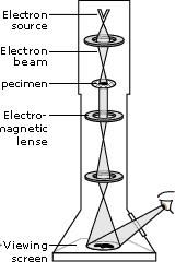
As the name implies, transmission electron microscopes ‘transmit’ electrons through thinly sliced specimens, providing enough resolving power to view structures as small as 0.05 nanometers (or 400 times more powerful than the strongest light microscope!).
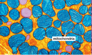
3) Scanning Electron Microscope
Similar to the Transmission Electron Microscope, the Scanning Electron Microscope (SEM) also uses electron beams to image small structures, however, rather than transmitting the electrons through a thinly sliced specimen, in SEM, the electron beams are reflected off of the surface of objects. The result is an image that has 3-dimensional depth.
Scanning Electron Microscopes are not quite as powerful as Transmission Electron Microscopes, have a maximum resolving power of around 1.5 nanometers (or about 13 times more powerful than the light microscope.


From: http://tysontrepidations.wordpress.com/2011/07/02/say-cheese-for-scanning-electron-microscopy/