Cytoskeleton
Introduction
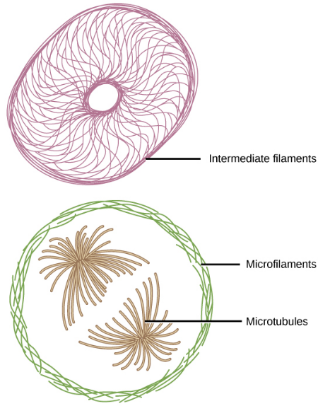
If you were to remove all the organelles from a cell, would the plasma membrane and the cytoplasm be the only components left? No. Within the cytoplasm, there would still be ions and organic molecules, plus a network of protein fibers that help maintain the cell’s shape, secure some organelles in specific positions, allow cytoplasm and vesicles to move within the cell, and enable cells within multicellular organisms to move. Collectively, scientists call this network of protein fibers the cytoskeleton. There are three types of fibers within the cytoskeleton: microfilaments, intermediate filaments, and microtubules. Here, we will examine each.
Learning
Microfilaments
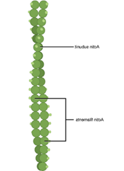
Of the three types of protein fibers in the cytoskeleton, microfilaments are the narrowest.
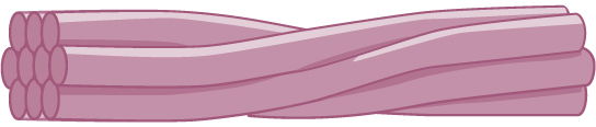
They function in cellular movement, composed of actin. For this reason, we they also call microfilaments actin filaments.
ATP powers actin to assemble, which serves as a track for movement. This enables the filament to engage in requiring motion, such as cell division in eukaryotic cells. Actin and myosin are plentiful in muscle cells. When your actin and other filaments called myosin slide past each other, your muscles contract.
Microfilaments also provide some rigidity and shape to the cell. They can depolymerize (disassemble) and reform quickly, thus enabling a cell to change its shape and move.
Intermediate Filaments
Several strands of fibrous proteins that are wound together comprise intermediate filaments. Cytoskeleton elements get their name from the fact that their diameter, 8 to 10 nm, is between those of microfilaments and microtubules.
Intermediate filaments consist of several intertwined strands of fibrous proteins and have no role in cell movement. Their function is purely structural.
The intermediate filaments are the most diverse group of cytoskeletal elements. Several fibrous protein types are in the intermediate filaments. You are probably most familiar with keratin, the fibrous protein that strengthens your hair, nails, and the skin’s epidermis.
Microtubules
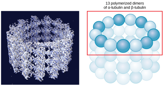
As their name implies, microtubules are small hollow tubes. Polymerized dimers of α-tubulin and β-tubulin, two globular proteins, comprise the microtubule’s walls. Microtubules are cytoskeletons’ widest components. They help the cell resist compression, provide a track along which vesicles move through the cell, and pull replicated chromosomes to opposite ends of a dividing cell. Like microfilaments, microtubules can disassemble and reform quickly.
Summary
The three types of filaments that make the cytoskeleton are:
- Microfilaments are the narrowest and function in cellular movement
- Intermediate filaments have no role in cell movement, their function is purely structural.
- Microtubules provide a track along which vesicles move through the cell
Sources:
“Cytoskeleton.” By OpenStax Biology 2e. Retrieved from: https://openstax.org/books/biology-2e/pages/4-5-the-cytoskeleton Licensed under: CC-BY: Attribution