Mitosis
Introduction
The central event of mitosis is the equal distribution of the parent cell’s DNA to two resulting nuclei, each destined for a progeny cell.
Furthermore, events which occur at the end of mitosis coincide with the start of cytokinesis, which involves the physical separation of the two progeny cells from the single parent cell.
In this module, we will explore the events which occur during mitosis and cytokinesis in more detail…
Learning
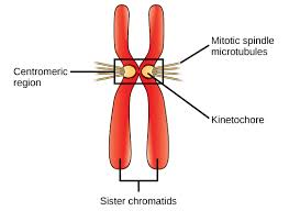
As mitosis begins, the nucleus of a typical eukaryotic cell contains twice the usual amount of DNA since each chromosome would have been replicated during the S-stage. These replicated chromosomes remain attached together after duplication, held together at a constriction point called the centromere.
The process of mitosis can be conceptualized as a series of four stages, each of which features easily identifiable events that are visible under the light microscope:
- Prophase
- Metaphase
- Anaphase
- Telophase & Cytokinesis
Prophase
The first stage of mitosis, called prophase (from the Greek terms pro, “before”; phase, “appearance”), is characterized by 4 key events:
- The appearance of visible, condensed chromosomes
- Condensed chromosomes are easier to transport (think of trying to throw a long piece of string across the room – this is much easier to do if the string is bundled into a compact ball!)
- Formation of ‘spindle microtubules’
- Structural proteins that work to distribute and transport cellular material, including the chromosomes, during cell division.
- Spindle microtubules attach to anchors at either end of the cell (called ‘poles’) and grow inward to create a ‘spindle’.
- Breakdown of nuclear envelope
- To distribute chromosomes to the progeny (daughter) cells, they must be freed from the nucleus.
- Attachment of spindle microtubules to the condensed chromosomes
- The spindle microtubules attached to the spindle ‘poles’ at one end grow inward and attach to the centromere region of the chromosomes. This attachment point on the chromosome is called the kinetochore.
Metaphase
After prophase, each chromosome is attached to the spindle pole by a spindle microtubule. The spindle microtubules begin to ‘pull’ on the chromosomes.
This force causes the chromosomes to line up in the middle of the cell, creating a visible structure when viewed under the microscope called the ‘metaphase plate’.

Anaphase

During the next stage of mitosis, called anaphase, the sister chromatids separate. Once they are separated, each chromatid is considered a complete, separate daughter chromosome.
The continued shortening of the spindle microtubules pulls the newly separated daughter chromosomes toward opposite spindle poles of the cell. This event results in the equal segregation of chromosomes into the two daughter cells.
Telophase
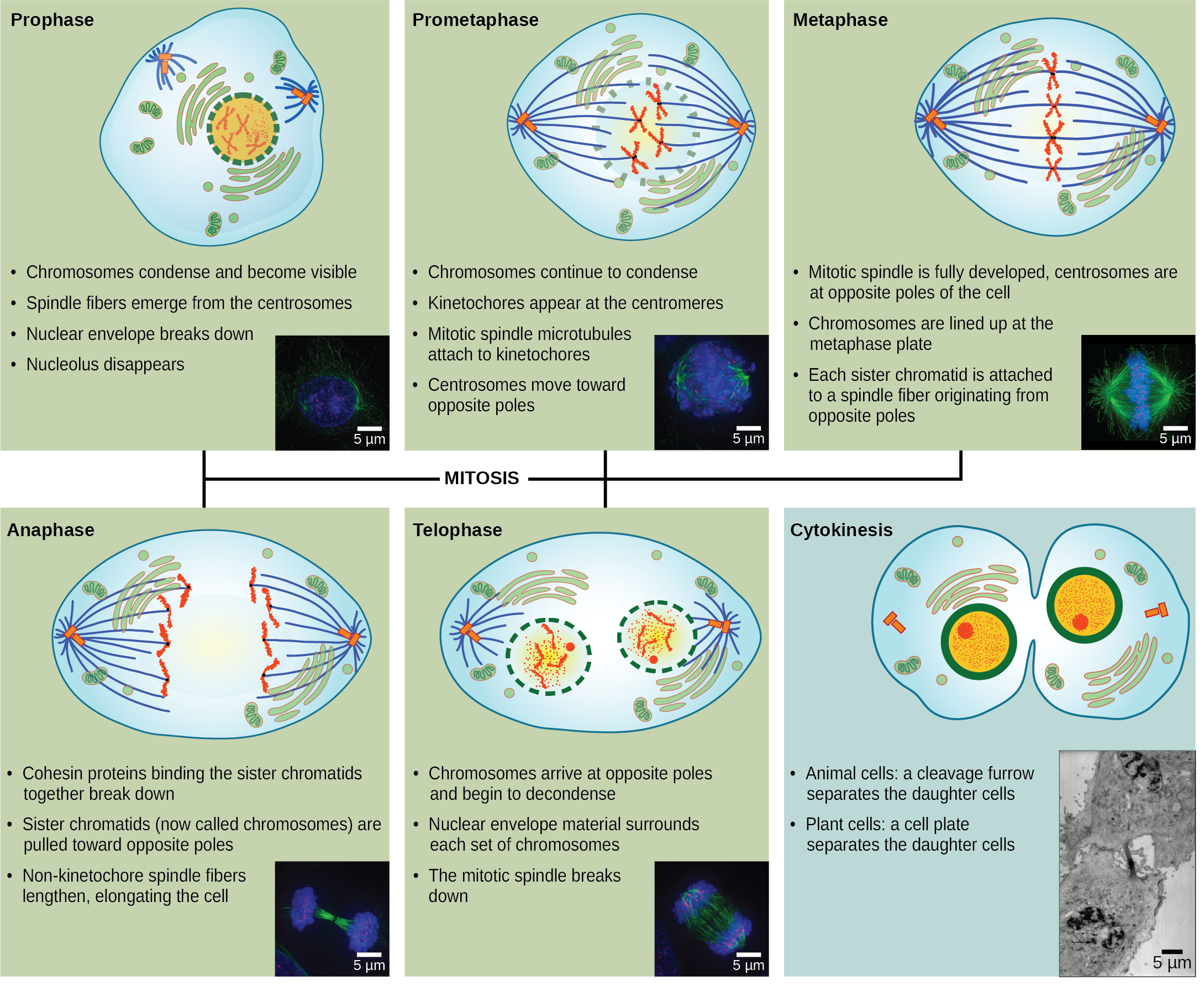
Telophase can be considered like the reverse of Prophase. In this sense, the following events occur:
- The daughter chromosomes are ‘de-condensed’.
- The ‘spindle microtubules’ are broken down.
- The nuclear envelope reforms around the newly segregated daughter chromosomes
Cytokinesis
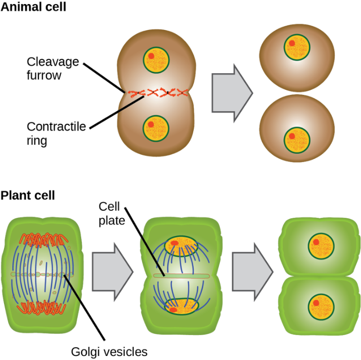
Telophase is followed by cytokinesis where the contents of the cell are physically divided into two as we saw in the Cell Cycle discussion.
Summary
Mitosis can be conceptualized as a series of four stages followed by cytokinesis which separates the parent cell into two identical progeny (daughter) cells, each containing a complete set of genetic information (chromosomes). The four stages of mitosis are:
- Prophase
- Metaphase
- Anaphase
- Telophase
Sources:
“Chapter 10 Cell Reproduction.” By OpenStax Biology 2e. Retrieved from: https:// https://openstax.org/details/books/biology-2e?Instructor%20resources/ Licensed under: CC-By: Attribution