Meiotic Cell Division
Introduction
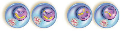
Meiotic cell division occurs exclusively in reproductive cells and helps create genetic diversity in the next generation of organisms. It starts with an initial chromosome crossing over (for genetic diversity, called Synapsis) followed by TWO rounds of specialized cell division – termed Meiosis I and Meiosis II. In this section, we will review the sequence of events that occur during meiotic cell division. For the purposes of comparison, we will also contrast how each event in meiosis differs from the events that occur during mitosis.
Learning
Meiosis I
The process of Meiosis I consists of a series of phases during which the following events occur. For the purpose of simplicity, our example will utilize a cell with only 2 pairs of chromosomes.
Prophase I
- Similarities with Mitosis
- Chromosomes condense
- Nuclear Envelope breaks down
- Meiotic Spindle forms
- Differences with Mitosis
- Chromosomes are not exact copies but have gone through “crossing over” to increase genetic diversity
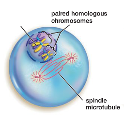
- Each duplicated, homologous pair of chromosomes will join together, forming a structure containing 4 chromosome strands of DNA called a ‘tetrad’
Metaphase I
- Similarities with Mitosis
- Spindle microtubules pull on chromosomes, causing them to line up along metaphase plate
- Differences with Mitosis
- Instead of sister chromatids lining up at metaphase plate, it is the tetrads that align in the middle of the cell.
- Important: the orientation of the tetrad (which homologous chromosome points to which spindle pole) is completely random
Anaphase I
- Similarities with Mitosis
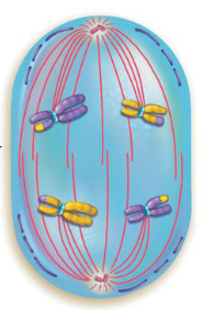
- Meiotic spindle pulls apart the chromosomes aligned at the center of the cell
- Differences with Mitosis
- Rather than the sister chromatids being separated, it is the tetrads which are pulled apart in Anaphase I
- Each member of the homologous pair will begin to move toward opposite spindle poles
Telophase I
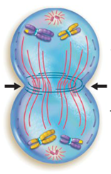
- Similarities with Mitosis
- Spindle microtubules start to disassemble.
- Differences with Mitosis
- Chromosomes do not de-condense
- Nuclear envelope does not re-form
- Each pole contains only one member of the pair of homologous chromosome(s).
- Note: Since there is only one version of each chromosome (even though they still exist as duplicated sister chromatids) at each pole, these opposite ends are ‘haploid’.
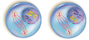
- Cytokinesis
- Cytokinesis will occur at the end of Meiosis I, resulting in two daughter cells, each haploid, but containing two copies of each chromosome (sister chromatids).
Meiosis II
Will typically occur immediately after the conclusion of Meiosis I. In Meiosis II, the sister chromatids will be separated following a series of steps that are almost identical to those which occur during mitosis.
Prophase II
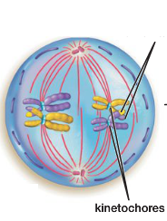
- Spindle microtubules re-form to attach to sister chromatids at the kinetochore
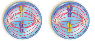
Metaphase II
- Spindle microtubules pull on the sister chromatids from each spindle pole
- Sister chromatids are aligned in the center of the cell along the metaphase plate
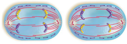
Anaphase II
- The sister chromatids are pulled apart toward opposite spindle poles, resulting in an individual daughter chromosome arriving at each pole
Telophase II
- The chromosomes will de-condense
- The nuclear envelope will reform at each spindle pole
- The spindle microtubules will disassemble
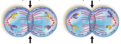
Cytokinesis
Cytokinesis will commence, splitting the cells into (4) haploid daughter cells, each containing only one member of the homologous chromosomes found in the original parent cell.
Summary
To review, meiotic cell division occurs exclusively in reproductive cells to create genetic diversity and consists of TWO rounds of cell division – termed Meiosis I and Meiosis II. The events that occur in Meiosis I and II result in the generation of (4) haploid gamete cells, each containing one set of chromosomes.